Unexpectedly, a fraction of green fluorescent CCL13 cells transfected with the promoterlesspRGLuc vector also expresses cryptic FLuc transcripts. Northern blot analysis of poly(A)+ RNA isolated from these cells gives diffuse signals instead of expected discrete bands.
Biological Significance
mRNA-LNPs encapsulating FLuc mRNA were physically labeled with the cyanine-5 dye DiR and intravenously injected into mice. The luciferase signal was localized mainly in the liver. This was because the LNPs were disseminated throughout the animal body and not only at the injection site, as with liposome-based mRNA. In contrast, an injection of naked mRNA did not result in solid gene expression at the injection site but at much more distant sites such as the muscle and the spleen.
Moreover, Northern blot analysis of poly(A)+ RNA isolated from cells transfected with pRGLuc and its promoterless variant pRG(-P)Luc showed a diffuse banding pattern that likely corresponds to several transcription initiation sites within the FLuc coding region in both samples. By contrast, a probe complementary to the 3′-end of the FLuc coding sequence yielded only a single signal in both samples, which indicates that only a limited number of cryptic transcripts were translated to luciferase activity.
What is FLuc mRNA contains the codon-optimized nucleotide sequence of the firefly luciferase protein from the Photinus pyralis. This mRNA is produced with capping technology, which results in a highly structured mRNA that mimics a fully processed natural mRNA. Furthermore, the mRNA is capped using an advanced capping system that utilizes the cap structure and has a uniform 100 bp poly-A tail. The mRNA is also dithiothyronine-treated to reduce antiviral responses and chemically modified to increase stability.
Transfection Efficiency
The FLuc mRNA was encapsulated into DiR-labeled LNPs for the study, and their biodistribution and protein expression in cells was monitored. The mRNA was efficiently released from the particles and was rapidly expressed in tissues. The mRNA expression in the liver was remarkably rapid and responsive.
It was analyzed with real-time RT-PCR to evaluate the transcriptional activity borne by the Fluc mRNA. PCR amplicons spanning the entire coding region showed that the transcription initiation site is located at the 5′ end of the transcript. Northern blot analysis confirmed this observation by showing a diffuse signal in the pRGLuc sample.
An intact CMV immediate-early promoter drives the transcription of the EGFP mRNA. In contrast, the cryptic promoter in the pFG vector shows only a weak activity compared to the solid immediate-early CMV promoter. This is most likely due to the different reading phases of the two genes, resulting in shorter transcripts with lower translation efficiency.
To assess the mRNA translatability of these new plasmids, they were co-transfected with a luciferase reporter gene in human cells.
Targeted Delivery
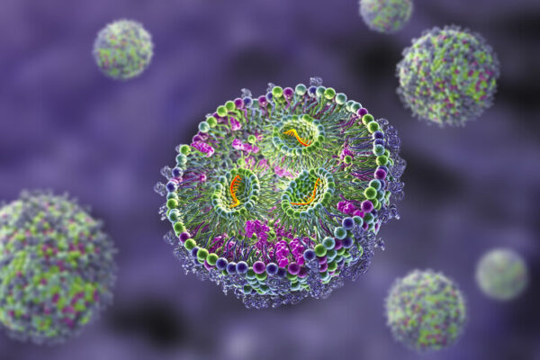
The FLuc mRNA is encapsulated in lipid nanoparticles (LNP) and can be delivered to cells or expressed in vivo. The mRNA is capped. Compared with uncapped mRNA, this LNP-encapsulated mRNA is more resistant to endosomal escape and cytoplasmic degradation.
The luciferase activity of the mRNA is determined in HEK293T cells. A luciferase activity significantly higher than that of the uncapped mRNA was observed. This result suggests that the mRNA’s cryptic promoter can drive transcription.
Northern blot analysis is performed using an ssRNA probe complementary to the 3′ half of the FLuc coding region to confirm this finding further. The ssRNA hybridized to the mRNA fragments isolated from CCL13 cells transfected with pRGLuc and a promoterless variant of pRGLuc, confirming that mRNA transcripts containing only the FLuc coding region are generated in the cell culture.
To investigate the biological effect of the cryptic promoter, the Fluc mRNA was encapsulated in various sizes of lipid nanoparticles and administered to mice by intramuscular injection. Injection of small-sized LNPs resulted in luciferase expression concentrated at the injection site, whereas larger particles were primarily released in the liver. In contrast, the medium-sized LNPs exhibited a balanced distribution of luciferase expression between the liver and injection site.
Analysis
Northern and real-time qRT-PCR assays analyzed the luciferase coding sequence of the FLuc mRNA. The results showed a gradual increase of transcripts complementary to the FLuc CDS in cells transfected with pRG and its promoterless variant (pRG(-P)Luc). The result suggests that transcription starts at several sites along the FLuc mRNA but that aberrant transcripts are subject to intensive degradation.
The authors also used the FLuc mRNA to investigate the lipoplexes’ biodistribution, sustainability, and organ-targeting characteristics following subcutaneous or intravenous administration. When plug mRNA was encapsulated in the MC3, KC2, or L319 LNPs and injected subcutaneously, the luciferase signal disappeared within two hours post-injection, consistent with rapid mRNA degradation. However, when plus mRNA was injected intravenously and delivered to the liver, luciferase signals were detectable 48 hours post-injection.
The authors developed a new split reporter strategy to visualize protein-protein interactions in vivo, where a reporter gene, such as FLuc, is dissected into two fragments fused to a pair of proteins that strongly interact with each other and produce a strong fusion product. They then use this fusion to generate labeled mRNA-LNPs and inject them into mice subcutaneously or intravenously.
